

Computer-Aided Drug Design Tutorials: 3.1. Visualization of Macromolecules
Protein Data Bank
The main database for protein structures is the Protein Data Bank, also known as the PDB, and its address is www.rcsb.org. Each structure in the Protein Data Bank has a unique 4-character alphanumeric identifier known as the PDB ID. You can retrieve all related structures using search by keyword, or access a particular structure by its PDB ID. Some molecular modeling programs, such as PyMOL and Chimera can directly retrieve structure files from the PDB.
The Research Collaboratory for Structural Bioinformatics, which manages the PDB, has created web-based tools that make it quite easy to search and understand the content of PDB files. Some of these tools require that Java Software is installed and that the Java plugin is enabled in your browser. Follow these steps to learn about some of these tools.
- Use local web browser or log into drug3 and open the RCSB PDB site in the web browser by typing mozilla www.rcsb.org
- Search the PDB by keyword oseltamivir to find all protein structures that contain the influenza drug Tamiflu. Brief information about each is shown. Select the one with the best resolution (smallest value) by clicking on its title line.
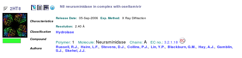
- Save the PDB file in your directory on the workstation as 2HT8.pdb by clicking on the "download" icon next to the PDB ID code.
- Visually examine the content of the PDB file by clicking on the "display text" icon. Scroll through this large file and notice that the structure contains only one polypeptide (chain identifier for all ATOMs is A) and one ligand, which has reside name G39. Close the window with text representation of the PDB file.
- Examine the information about this structure. Notice various icons within the description that link out to external resources. For example, the M icon in the Primary Citation field leads to the MEDLINE entry for this publication, allowing to read the abstract, and possibly access the original research publications.
- The Chemical Component field is one of the most interesting from the drug design viewpoint. It provides information about every molecule that is bound to the protein. Clicking on the Drug Similarity [View] icons will lead out from PDB to a Super Drug Database tool that finds well-known molecules that are most similar to this molecule. Clicking on the [View] under Ligand Interaction will open a Java application that analyzes the structure of the binding pocket. When you click on the [View] under Ligand Structure, you can see the full name, SMILES string and the chemical structure of the molecule.
- Click on the G39 in the Chemical Component field to see all the protein structures where this ligand is present. Notice that this list is longer than your original "oseltamivir" search because the molecule was not called oseltamivir during the early periods of its study.
- Click on the External Links to see a rather long list of tools. One that might be useful for us is the PDBsum. Follow this link and briefly examine the views under Protein tab, under the Ligand tab, and under the Clefts tab. Notice how the LigPlot image provides a 2D view of the active site interactions and how you can identify possible binding sites in the Clefts tab. Close the PDBsum window.
- Click on the External Links and select Computed Atlas of Surface Topography of proteins (CASTp). The CASTp server allows identification and characterization of pockets and cavities in protein structures. In the CASTp window, select Calculation Request, then enter 2ht8 or upload the structure that you had saved earlier. Examine different pockets in the structure by checking the largest pockets from the left-side menu. Close the CASTp server window when done.
- You may close the web browser on the workstation and follow to the next part of the tutorial using a browser on the SGI computer.