

HIV protease is a dimeric enzyme. The structure you just analyzed was only one monomer of the dimer. Many proteins function as dimers or higher oligomers.
To download the structure of the dimeric enzyme, search the PDB Data Bank for the code '7HVP' and download the file. The structure contains a non-hydrolyzable peptide inhibitor different from the previous inhibitor. Open 7HVP.pdb with SYBYL after deleting the previous monomer.
As frequently the is case, the structure on the screen is quite complex. Even experienced researchers find it difficult to analyze such structures without some visual signposts. A simple, yet powerful trick is to use unique colors to highlight important parts of the molecule. For example, you can easily visualize the bound inhibitor if all its atoms are colored green. To select and color atoms of the inhibitor, follow the instructions below:
In the 'View' menu, under Color select 'Atoms '
Click on the button marked Substructures, select ( these are at the very bottom!)
ACE0, SER1, LEU2, ASN3, PHE4, CH25, PRO6, ILE7, VAL8, OME9, and click OK
Another window pops up; select GREEN as the color of the inhibitor peptide and hit OK
Put on stereoscopic glasses and hit F7 to bring up the stereo display. See how the inhibitor fits into the active site. Identify at least one amino acid of the protein that is very close to the inhibitor. You can do this as follows:
In the 'View' menu, under Label select 'Substructure '
Click on any atom an amino acid in the HIV protease that is near the inhibitor.
The name and sequence number of this amino acid are displayed. Write it down in your notebook
Medicinal chemists can design new HIV drugs by carefully studying how active site residues of the HIV protease interact with bound inhibitors. HIV, however, is capable of changing its active site residues so that drugs will not bind to the mutant protein. This causes drug-resistant strains to appear quite quickly, and new drugs must be developed to ward off the the mutant virus. Drug resistance is common in many diseases, but particularly serious for HIV infections.
The scene is still quite busy and we can simplify the picture by showing only the cartoon of the protein backbone. We again will use the Ribbon/Tube model of the backbone:
In the 'Biopolymer' menu, under Display select 'Ribbon/Tube '
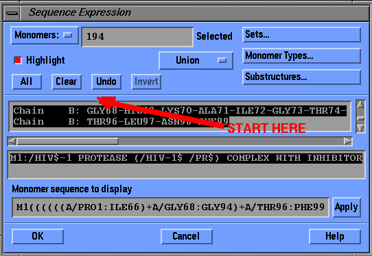
Highlight Chains A and B by dragging cursor over the sequence window and hit OK
Another window pops up; answer OK to color the protein according to the secondary structure.
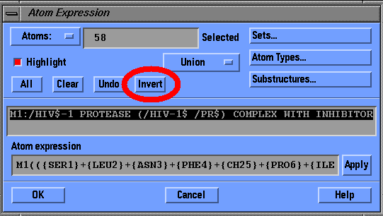
In the 'View' select 'Undisplay Atoms...'
Click on the button marked Substructures, select (these are at the very bottom!)
ACE0, SER1, LEU2, ASN3, PHE4, CH25, PRO6, ILE7, VAL8, OME9, and click OK
Click Invert to select everything but the inhibitor peptide, and hit OK
Can you identify the dimeric structure of the protein? Also, note that there is a defect in the rendering of this dimer: two yellow arrowheads are not connected! This is because the structure of highly flexible parts of the protein often cannot be determined from X-ray diffraction data.
Studies have shown that the activity of HIV protease depends on the active site aspartic acid (Asp25) residue. To selectively show this residue, follow these steps:
Assignments (HIV Protease continues):In 'View', select 'Display Atoms...'
Click on the button marked Substructures
Select ASP25 ... ASP25 (There are two Asp25 residues, as this is a dimer!)
Click OK and OK to finish selection
The structure and function of HIV protease are further described in a Chime-based tutorial from University of Virginia in Charlottesville. Please note that you need a Chime plug-in to see this structure. The plug-in is not installed on SGI computers.